World’s oldest heart preserved in 380 million-year-old armored fish
A team of Australian scientists has discovered the world’s oldest heart, part of the fossilized remains of an armored fish that died some 380 million years ago. The fish also had a fossilized stomach, liver, and intestine.
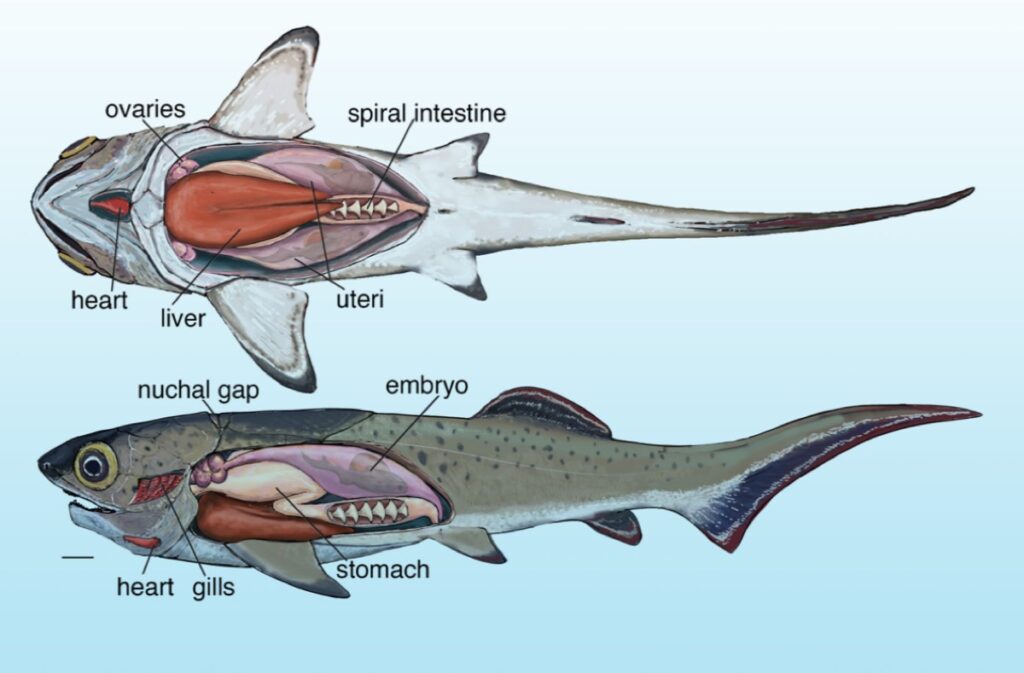
All the organs were arranged much like similar organs in modern shark anatomy, according to a recent paper published in the journal Science.
As we’ve reported previously, most fossils are bone, shells, teeth, and other forms of “hard” tissue, but occasionally fossils are discovered that preserve soft tissues like skin, muscles, organs—or even the occasional eyeball. This can tell scientists much about aspects of the biology, ecology, and evolution of such ancient organisms that skeletons alone can’t convey.
For instance, earlier this year, researchers created a highly detailed 3D model of a 365 million-year-old ammonite fossil from the Jurassic period by combining advanced imaging techniques, revealing internal muscles that had never been previously observed. Among other findings, the researchers observed paired muscles extending from the ammonite’s body, which they surmise the animal used to retract itself further into its shell to avoid predators.
And last month, British researchers described their experiments monitoring dead sea bass carcasses as they rotted over the course of 70 days to gain insights into how (and why) the soft tissues of internal organs can be selectively preserved in the fossil record.
One of the best ways that soft tissue can turn into rock is when it is replaced by a mineral called calcium phosphate (sometimes called apatite). Specifically, muscles, stomachs, and intestines tend to “phosphatize” much more frequently than other organs like kidneys and gonads. The authors concluded that the phosphorus content of specific organ tissue contributes to this unusual selection bias for which soft tissues are preserved in the fossil record.
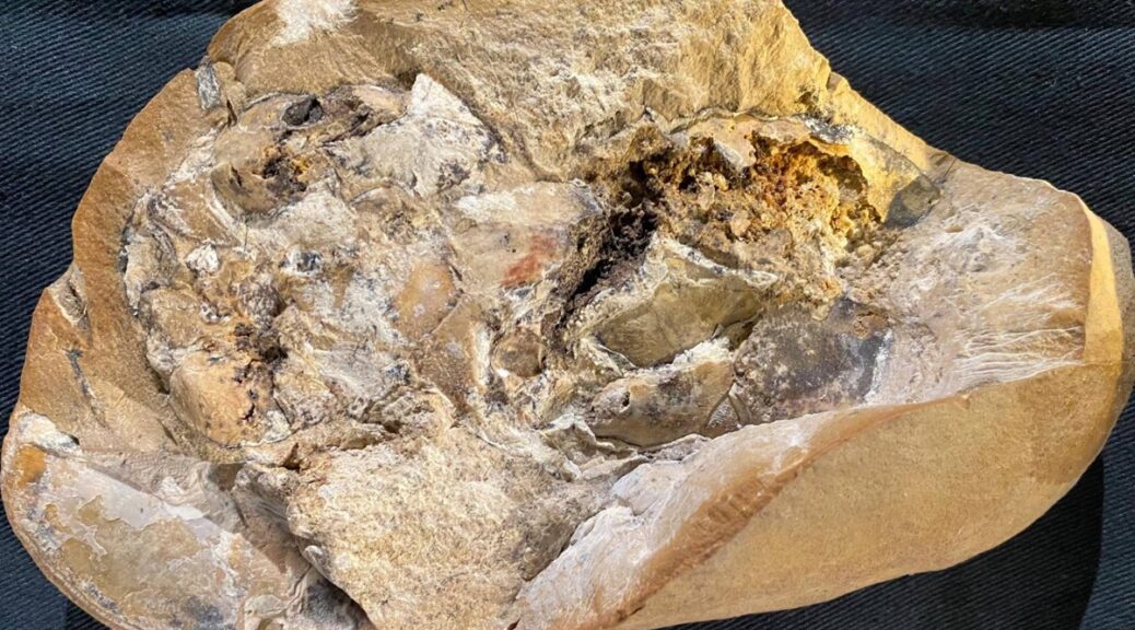
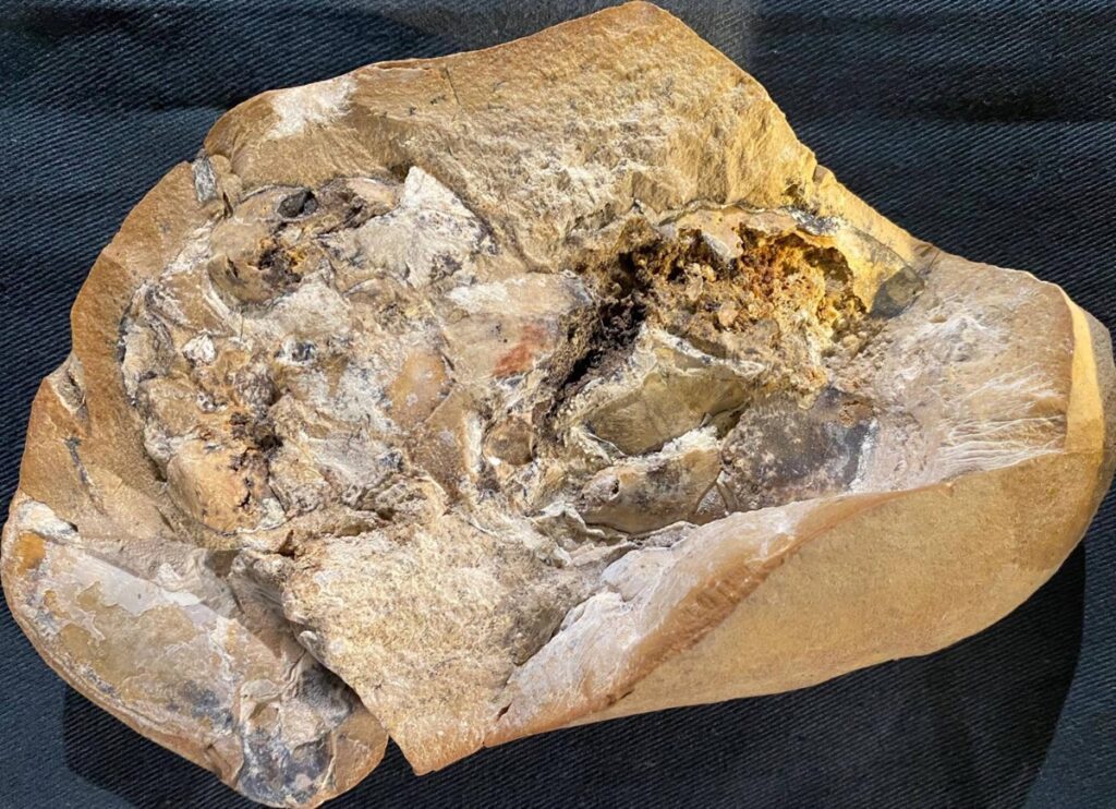
The fossilized specimens examined in this latest paper were collected from the Gogo Formation in Western Australia, which was once a reef and is rich in exceptionally well-preserved Devonian fossils, such as the class of armored prehistoric fish known as placoderms. That preservation includes soft tissues, including nerves.
In 2005, paleontologists even excavated a new species of placoderm, dubbed Materpiscis (“mother fish”), with an embryo still attached by an umbilical cord—evidence that at least some species of armored fish gave birth to well-developed live offspring.
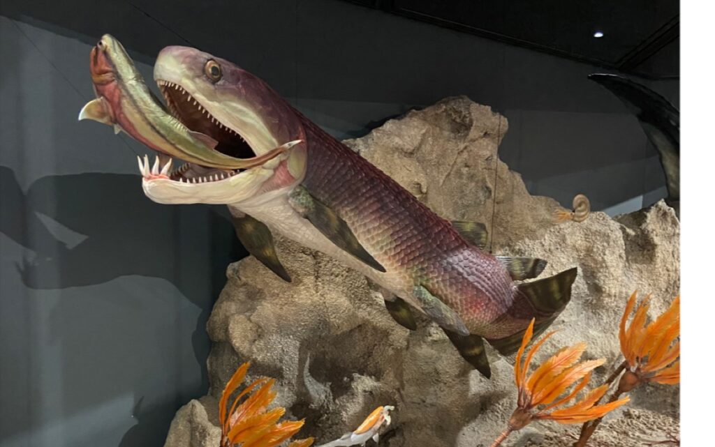
According to the authors of this latest paper, placoderms were among the earliest jawed vertebrates, the evolution of which involved significant changes to skeletal structure and soft anatomy. Because the preservation of soft tissue is so rare in the fossil record, the samples collected at the Gogo Formation (and now housed in the public collections of the Western Australian Museum and the Museum of Victoria) could hold clues about how this transition occurred—specifically, how the head and neck region changed to accommodate jaws.
“What’s really exceptional about the Gogo fishes is that their soft tissues are preserved in three dimensions,” said co-author Per Ahlberg of Uppsala University.
“Most cases of soft-tissue preservation are found in flattened fossils, where the soft anatomy is little more than a stain on the rock. We are also very fortunate in that modern scanning techniques allow us to study these fragile soft tissues without destroying them. A couple of decades ago, the project would have been impossible.”
Paleontologists collected the samples by splitting limestone concretions in the field, then taping the broken pieces together for transport. The researchers were able to scan the intact samples using neutron beams and synchrotron radiation. Then, they constructed 3D images of the soft tissues preserved within based on the different densities of minerals deposited by bacteria and the surrounding matrix of rock.
The result: the first 3D model of a complex, flat s-shaped heart with two distinct chambers. The team also imaged a thick-walled stomach with intact intestines and a liver, separated from the heart; they also noted the absence of lungs. The fossilized liver was quite large and likely helped the fish stay buoyant, per the authors. It’s the first time scientists have been able to see the arrangement of organs inside a primitive jawed fish.
“As a paleontologist who has studied fossils for more than 20 years, I was truly amazed to find a 3D and beautifully preserved heart in a 380 million-year-old ancestor,” said co-author Kate Trinajstic, a vertebrate paleontologist at Curtin University.
“Evolution is often thought of as a series of small steps, but these ancient fossils suggest there was a larger leap between jawless and jawed vertebrates. These fish literally have their hearts in their mouths and under their gills—just like sharks today.”